
Interventional Oncology
Quality improvement
IR Quarterly
Radiation exposure is an unavoidable part of interventional radiology, but while the risks are understood, they’re not always top of mind. Unlike other occupational hazards, radiation does not announce itself in the moment. Its effects accumulate quietly over time, making vigilance both essential and difficult to sustain.
“I think all IRs are students of radiation safety. You kind of need to be actively engaged in radiation safety to be in this field,” said John Fritz Angle, MD, FSIR, who has been involved in developing multiple radiation safety papers and guidelines.
The challenge, however, lies not just in awareness of the risks but in maintaining consistent safety behaviors and continuous vigilance.
“It’s an invisible harm because you don’t see the radiation damaging you until it’s too late,” said Nikki Keefe, MD. Dr. Keefe, along with Dr. Angle and Kyle Jones, PhD, FSIR, submitted a response to the American College of Radiology’s recent Blue Ribbon Panel on Fluoroscopy Safety.
Solutions to unnecessary radiation exposure exist, Dr. Keefe and Dr. Angle say, but they require behavioral, technological and institutional alignment. It’s a burden that should not fall solely on individual operators; rather, it should become a shared responsibility embedded in the culture of IR and medicine in general.
As leaders in the field of radiation safety, IRs are uniquely positioned to ensure proper training and protection for themselves and their colleagues—but every IR should be well versed in safety practices and advocate for their own health.
Efficiency vs. safety
In day-to-day practice, radiation safety often competes with efficiency.
“There is so much pressure to get things done quickly, and most radiation protection practice takes time,” said Dr. Angle. “There is an intrinsic push to prioritize the patient and the caseload, and as a result radiation safety doesn’t always get the attention it deserves.”
This tension can lead to subtle but significant behavioral drift over time. Even experienced and well-trained physicians are not immune.
“I definitely fall into complacency at times,” Dr. Angle said. “It’s something for us all to be cognizant of.”
Dr. Keefe echoed the same concern from another perspective, noting that habits—good or bad—are often shaped by environment and observation.
“People become laissez-faire about their exposure. Sometimes they’re not properly trained in radiation safety, or they forget about it over time, or they think they’re impervious to it,” she said. “It’s easy to fall into the pattern of, ‘this is what everybody else does,’ and not think about it.”
This is why it is crucial to start safety training from the very beginning, when new IRs are learning the basics of practice—and inadvertently being exposed to higher rates than their colleagues.
“Trainees may need to do extra runs or take longer in an area while learning, and so their radiation dose is invariably going to be higher than that of attendings,” Dr. Keefe said. “So as program directors, we need to prioritize teaching our younger generations how to protect themselves from day one.”
Making the most of dosimeters
SIR and the Cardiovascular and Interventional Radiological Society of Europe (CIRSE) recently published an updated occupational safety guideline which recommends best practices for tracking exposure.
As described in the guideline, the International Commission on Radiological Protection (ICRP) and National Council on Radiation Protection and Measurements (NCRP) recommend that interventionalists use the two-dosimeter method. This approach allows for a better estimate of whole-body exposure than a single reading.
“The issue, though, is that dosimetry methodology is still not standardized across all institutions and even varies from region to region,” Dr. Angle said. Accepted safety limits differ between countries, as do best practices for how many dosimetry devices to use and what kind. At many institutions, a single dosimeter is still considered standard; Dr. Keefe said she has used the two-dosimeter approach only while pregnant.
But safety technology is evolving.
“At our institution, we have access to an instantaneous dose display,” Dr. Angle said. “It’s a second badge that I can wear, and we hang the screen next to our fluoroscopy screen, which gives us moment-to-moment dosimetry.”
Having access to this technology has been a valuable learning experience, Dr. Angle said. The system provides immediate feedback on exposure, allowing operators to connect specific actions with measurable dose changes.
“Our exposure is extremely episodic, and so certain decisions make a huge difference in our cumulative dose,” Dr. Angle said. For example, seemingly minor choices—such as whether to step out of the room or adjust detector angle—can meaningfully reduce exposure. Dr. Keefe described similar insights from her experience using real-time dosimetry during training.
“It was astounding the information that you gathered as to where was the safest to stand,” she said. “It was very interesting to see the effects of doing a DSA versus fluoroscopy and how substantial those differences were. Experiencing real-time feedback allows you to appreciate radiation effects at different understanding than reading a paper. Even something such as where you moved in a room, or whether you stepped out during a DSA could make a huge impact on your dosage.”
Still, adoption is limited. Cost and lack of standardization mean these technologies are not universally available, leaving many practitioners without this real-time learning opportunity.
Small changes, significant impact
While advanced tools offer valuable insight, many of the most effective safety strategies are simple, low-cost and immediately actionable.
“There’s a lot that we can do that’s very easy,” Dr. Keefe said, such as narrowing cone beams, minimizing magnification, increasing distance from the source and properly using shielding. IRs can also minimize the amount of digital subtraction angiography they perform or step out of the room, as clinically appropriate.
She also reminds IRs that they don’t need to stand right next to the fluoroscopy machine; while completing procedures such as dialysis work, IRs can utilize extension tubing to give themselves more space.
“People underestimate how much impact it can have if they simply leave the room. If you stand in the corner during a cone beam, you still get a fair amount of radiation,” said Dr. Angle. “It takes more time, yes, but you can always just leave the room.”
The pregnancy gap
Radiation safety becomes even more complex when applied to specific populations, particularly pregnant IRs.
Dr. Keefe became pregnant while she was a senior resident and again as a junior attending and found very little information on pregnancy and radiation safety from an IR perspective.
“My program had never had a pregnant IR fellow before,” she said. “Previously, if any trainees were pregnant, they just didn’t do IR at that time. Obviously, that’s not an option if you’re a fellow.”
As a result, Dr. Keefe conducted her own research and interviews, which has since led her to become extremely passionate about radiation safety.
The lack of guidance has led to many misconceptions, Dr. Keefe said.
“Some people think it’s completely unsafe to be pregnant and practice IR, but that’s not true, as long as you take proper precautions.”
This includes using two dosimeters—one in the standard collar placement and another beneath the lead—and being familiar with recommended dose guidelines. Contrary to some assumptions, it does not mean wearing more lead.
“Adding more weight can actually introduce new risks and musculoskeletal considerations,” Dr. Keefe said. The weight of protective equipment already has orthopedic impacts on long-term users, and adding more can be dangerous. 0.5 mm Pb equivalent is recommended which is what typical physician lead thickness is anyways.
The hidden cost of protection
“There's no question that radiation protective gear leads to serious spine problems for some operators,” Dr. Angle said.
Dr. Angle, Dr. Keefe and Dr. Jones recently published a commentary on the findings from The Society for Cardiovascular Angiography and Interventions (SCAI)’s summit on radiation and orthopedic risks in fluoroscopic labs. The summit’s findings included substantial discussion of the orthopedic costs of extended lead use, such as spine pain and degenerative disease.
Panelists also discussed the benefits of enhanced radiation protection devices (ERPDs), which may provide suitable protection while eliminating or reducing the need for lead aprons.
However, ERPDs are not practical for all situations or procedures.
“IR presents a unique challenge given the wide variety of procedures performed and variable and often changing location where personnel stand during a single procedure. ERPDs require further investigation to confirm their suitability and effectiveness in IR practice environments,” wrote Dr. Keefe, Dr. Angle and Dr. Jones.
Despite the questions, Dr. Angle said he was pleasantly surprised by the attention paid to the orthopedic impact of practice—something he urges SIR and other IR leaders to investigate further.
System-level barriers
Though all IRs need to be aware of safety protocols, the responsibility should not fall entirely on each individual physician, Dr. Keefe said.
“In all radiation-facing fields, we are risking our own health for the patient,” she said. “All the things we can do to minimize our exposure come at a financial cost—but they don’t actually change patient outcomes.”
As a result, Dr. Keefe says, there is no financial incentive for institutions to invest in practitioner safety. This means that while technology may be advancing—such as radiation pads on patients to minimize dose, more ergonomic lead aprons, ERPDs or even real-time dosimeters—these tools are not often prioritized because they are an added expense without a clear return on investment.
In many places, IRs are even required to purchase their own lead, Dr. Keefe says, something she describes as the bare minimum of protection.
And as more specialties embrace minimally invasive, image-guided medicine, findings from these recent summits indicate that more and more physicians are being exposed to radiation without proper education. As a result, safety has become an individual burden, rather than a collective priority. As radiation exposure among different specialties becomes more prominent, IR’s can serve as advocates and educators in this area.
“The radiation-facing specialties, all need to get together and jointly advocate that this is unacceptable,” Dr. Keefe said. “We are putting our livelihood and our lives at risk. Yes, we all know what we signed up for, but much of this is preventable.”
From awareness to accountability
Radiation safety in interventional radiology is not a new concept. The risks are well documented and many solutions already exist. But closing the gap between knowledge and practice remains the central challenge.
“We can find a way to give radiation safety the time it deserves for every patient and every interventional radiologist,” Dr. Angle said.
Doing so will require more than individual awareness. It will demand a cultural shift—one that prioritizes safety alongside efficiency, leverages technology for education and pushes institutions to align incentives with practitioner well-being.
Resources:
- Miller D, Vano E, Balter S, et al. Occupational Protection in Interventional Radiology. A Joint Guideline of the Cardiovascular and Interventional Radiological Society of Europe and the Society of Interventional Radiology. J Vasc Interv Radiol. 2026; 37.
- Gress DA, et al. Recommendations From the Blue Ribbon Panel on Fluoroscopy Safety. J Am Coll Radiol. 2026 May;23(5):779-789.
- Angle J, Jones A, Keefe N. Commentary on Blue Ribbon Panel on Fluoroscopy Safety. J Vasc Interv Radiol. 2026 May 20;37(8):108859.
- Salavitabar, A, Vora A.N, Altschul D, et al. ALARA+: Summit on radiation and orthopedic risks in fluoroscopic laboratories: endorsed by ACC, ACVP, ASE, HRS, SCAI, SIR, SNIS, SVIN, and SVS. J Vasc Interv Radiol. 2026; 37:108689
- Angle J, Jones A, Keefe N. Commentary on “SCAI Summit on Radiation and Orthopedic Risks in Fluoroscopic Labs”. J Vasc Interv Radiol. 2026; 37.
Related
-
IRQ Clinical Practice
![]()
Minimizing delays from lung cancer diagnosis to treatment
![]()
Time between lung cancer diagnosis and treatment is a critical challenge in oncology care. Delay in treatment results in worse clinical outcomes. A new multidisciplinary model out of Dayton VA Medical Center has found a way to bridge the gaps.
-
IRQ Clinical Practice
![]()
Invisible risks, visible solutions
![]()
Advancing radiation safety in IR
Safety
All levels
Women in IR
-
IRQ Clinical Practice
![]()
New Category III codes for July 2026
![]()
Coding for histotripsy of malignant pancreatic tissue and surface mesh representations and applications.
Christina G. Marks, MD
Coding Q&A
Coding, billing and reimbursement
-
IRQ Clinical Practice
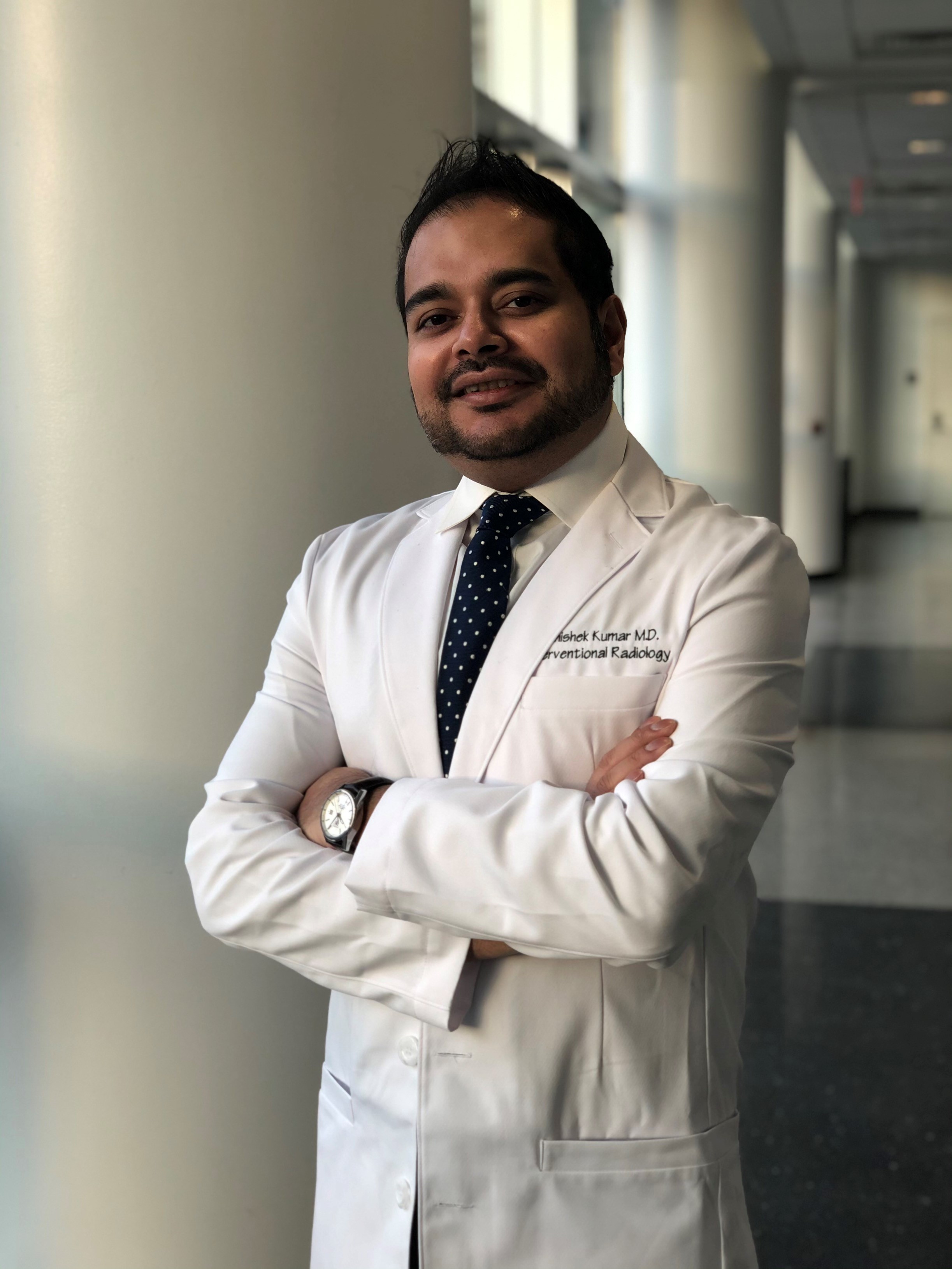
High-impact IR program building
Dr. Abhishek Kumar's Y-90 program has become the highest-volume radioembolization program in New Jersey. Here's how he did it.
Practice development
Practice profiles
-
IRQ Clinical Practice
![]()
Coding Q&A: Using a pressure-directed catheter for radioembolization of a hepatic tumor
![]()
Q: When an interventional radiologist uses a pressure-directed catheter for radioembolization of a hepatic tumor in the hospital outpatient setting, does this change the coding reported by the physician, i.e., does the physician report HCPCS (Healthcare Common Procedure Coding System) codes C1982 and C9797?
Coding Q&A
Christina G. Marks, MD
Coding, billing and reimbursement
-
IRQ Clinical Practice
![]()
E/M coding
![]()
Optimizing and capturing the full scope of IR
Economics
Coding, billing and reimbursement
Winter 2026
![]()
Related
![]()
-
IRQ Clinical Practice
Minimizing delays from lung cancer diagnosis to treatment
Time between lung cancer diagnosis and treatment is a critical challenge in oncology care. Delay in treatment results in worse clinical outcomes. A new multidisciplinary model out of Dayton VA Medical Center has found a way to bridge the gaps.
-
IRQ Clinical Practice
Invisible risks, visible solutions
Advancing radiation safety in IR
Safety All levels Women in IR -
IRQ Clinical Practice
New Category III codes for July 2026
Coding for histotripsy of malignant pancreatic tissue and surface mesh representations and applications.
Christina G. Marks, MD Coding Q&A Coding, billing and reimbursement -
IRQ Clinical Practice
High-impact IR program building
Dr. Abhishek Kumar's Y-90 program has become the highest-volume radioembolization program in New Jersey. Here's how he did it.
Practice development Practice profiles -
IRQ Clinical Practice
Coding Q&A: Using a pressure-directed catheter for radioembolization of a hepatic tumor
Q: When an interventional radiologist uses a pressure-directed catheter for radioembolization of a hepatic tumor in the hospital outpatient setting, does this change the coding reported by the physician, i.e., does the physician report HCPCS (Healthcare Common Procedure Coding System) codes C1982 and C9797?
Coding Q&A Christina G. Marks, MD Coding, billing and reimbursement -
IRQ Clinical Practice
E/M coding
Optimizing and capturing the full scope of IR
Economics Coding, billing and reimbursement Winter 2026
Related