
Inside Access
JVIR
Interventional Oncology
IR Quarterly
Minimizing delays from lung cancer diagnosis to treatment
BY ROBERT F. SHORT, MD, PHD
Lung cancer is the number one cause of cancer death and disproportionately affects U.S. veterans. The Southwest Ohio River Valley—the area serviced by Dayton Veterans Affairs Medical Center—has some of the highest lung cancer incidence in the country.
The Dayton VA Medical Center is uniquely positioned to handle this patient population. As part of our institution’s interventional radiology/interventional oncology program we have a busy lung biopsy and lung cancer image-guide thermal ablation (IGTA) program, alongside surgery and stereotactic body radiotherapy (SBRT). As a result, our institution is one of only a few high-volume centers in the U.S. to offer all three definitive lung cancer therapies.
However, time between lung cancer diagnosis and treatment is a critical challenge in oncology care. Delay in treatment results in worse clinical outcomes. To minimize time delays and improve patient care, Dayton VA Medical Center has launched a new multidisciplinary model called the Minimally Invasive Nodule Therapy (MINT) Clinic.

Impact of delays
In 2017, an article in the Federal Register utilized historical data1 from our institution which looked at the multipronged process that patients undergo from identification of a lung nodule through work-up, staging and treatment. The data from this paper spanned from 2011–2013—interestingly, just before the 2011 National Lung Screening Trial and the USPTF 2013 recommendation for annual low-dose CT lung cancer screening (LCS).
Data show that Veterans Health Administration institutes tends to outperform the private sector in terms of preventative medicine and screening2 and LCS is no different. In fact, the VHA immediately went to work when the USPTF released recommendations to adopt LCS across the VA system. After several pilot sites demonstrated success, the VA began a national rollout in 2017 for LCS programs with dedicated personnel and infrastructure. Our facility began steadily increasing enrollment in LCS as a result.
A growing demand for treatment
Around that same time, we began offering pulmonary ablation for select patients who were not surgical candidates. This coincided with an update to the National Cancer Center Network guidelines in 2018 which clarified the role for image-guided thermal ablation for pulmonary tumors. Our VA site then became a Lung Precision Oncology Program site as part of a national VHA oncology initiative to expand beyond screening to include patient-specific treatment, further expanding on the goals of the LCS program.
In 2021, the USPTF broadened screening eligibility (50–80 years old, > 20 pack years smoking), which substantially increased the volume of patients eligible for LCS. As a result, we began to see a lot more patients and identified substantially more potential cancers. These potential cancers then had to go through the post-imaging detection work-up—a process that had not changed much since 2013.
Essentially, screening had expanded dramatically, but our treatment pathways hadn’t.
Investigating the process map
The goals of the project were relatively simple: Get patients from identification of a suspicious lung nodule to initiation of definitive therapy. The clock starts ticking when a suspicious lung nodule is identified on LCS (generally a category 4B) and ends when the patient receives one of three definitive therapies offered at our institution: surgical resection, radiation (e.g. SBRT) or IGTA.
The process map for lung cancer is mostly defined clinically. When a suspicious nodule is identified, there are several steps in work-up that need to happen. The first is a biopsy. We knew we were seeing more—a lot more—lung biopsies and took steps to triage these patients for “fast tracking” to biopsy.
This is just one step. Patients also undergo pretreatment evaluation for possible lung cancer staging with a PET scan and, when indicated, mediastinal sampling, as well as assessing surgical candidacy with pulmonary function testing (PFT). It is then discussed at a multidisciplinary tumor committee followed by referral to an appropriate treating service.
Our process was no different than most centers where these often occur sequentially. We then broke down and examined each step to see where the greatest avoidable delays were occurring.
The testing bottleneck
We measured the time from referral to appointment and the impact that had on the overall length of time to therapy. One thing we noticed in the multidisciplinary tumor committee was that often after a biopsy, a patient would be presented, but the pre-treatment evaluation might not be complete because the patient was missing PFTs. The patient would then have to get PFTs and then potentially be re-submitted after the PFTs established upfront that the patient was a poor surgical candidate.
PFT is the biggest objective determinant of surgical candidacy. Some patients with poor PFTs might still be surgical candidates; however, these patients are more likely to be subject to lasting morbidity. We found that sometimes it took a month or more to get the PFTs done, plus in the rare instance of a post-biopsy pneumothorax PFTs would have to be delayed by 2 weeks or more. Thus, the patient’s PET scan might be out of date by that time (ideally the PET would be performed within 30 days of therapy for SBRT, according to the ACR; 60 days per NCCN).
Repeating the scan meant increased cost, patient inconvenience and delay. This sequential approach of siloed appointments could create significant delays, particularly for those patients who were not good surgical candidates, i.e. those who might ultimately be treated by SBRT or IGTA.
If one looks at the flowchart for the NCCN guidelines for a Stage IA NSCLC, one of the interesting things we noticed was the phrase “if not already performed” next to two key features of the patient work-up: the PET scan and PFT. A few other elements in the updated guidelines were important as well:
- For peripheral nodules < 3 cm with a negative PET scan, mediastinal sampling is optional, and
- If mediastinal sampling was desired, performance concurrent with surgical resection is preferable.
Taken together, these were the areas where most time was added between diagnosis and therapy. Solving these problems would take true multidisciplinary collaboration. Having completed preliminary investigations of our own, we formally engaged our hospital quality improvement team to help with the project.
Building a multidisciplinary team
From the onset, we looked to involve as many of the interested stakeholders in the process as we could. We shared our data to underscore the need for change and looked to generate enthusiasm along the way. We then created protocols that standardized our process and sought buy-in along the way through committee approvals. The enthusiasm from our Lung Cancer Screening coordinators for seeing their patients move through a suspicious nodule work-up to definitive therapy really put wind into the sails of the program.
Our multidisciplinary team included thoracic surgery, pulmonology, oncology, radiation oncology, and IR. Together, we were able to comprehensively review our institutional practices.
Finding the key pieces
First, we examined routine use of separated mediastinal staging procedures and the impact on time to therapy. After evaluating our data and considering updated guidelines, we collectively agreed that if PET scans could be obtained closer to therapeutic intervention decisions, it would obviate the need for most separate-procedure mediastinal evaluations (e.g. bronchoscopy, mediastinoscopy). Similarly, we noticed the time from referral to PFT was not what we hoped. In addition, the timing of the PET scan was essential to avoid the cost and delays associated with repeating it. That’s when we asked ourselves, “Why can’t these be ‘already performed’?”
Leveraging new technology
The eureka moment came when researching possibilities to expand the PFT capacity within our facility. We discovered that a cabinless PFT device had recently been devised and validated, published in Chest in 2021.3 We realized that this tabletop machine created an opportunity to redesign the workflow. Having achieved administrative buy-in via our quality team, we were then able to work with our hospital innovations team to rapidly acquire the device.
Training and utilizing staff
The next major advance was not in technology but in people: we could deploy the technology in the IR clinic by training our IR nurses to perform PFTs. Doing so would allow PFTs to be performed immediately prior to a biopsy for any patient that did not already have them. We saw this as analogous to performing EKGs in our department, a process familiar to many.
The IR nursing team, coordinators and our physician assistants were critical for not only owning the procedure, but the patient’s path. Now if a patient is referred for biopsy, we make sure those “if not already performed” elements (PET and PFTs) are not done, we get them done.
Combining the testing process
The quality team, IR nursing and the pulmonary service (including the physicians and respiratory therapists) worked together to devise a sanctioned process to provide for PFT performance in IR. Taking it a step further, we realized we could work with our colleagues within the therapeutic and diagnostic imaging department to coordinate with nuclear medicine to arrange pre-biopsy PET scans for the same day as the planned biopsy. Now, what was once three separate appointments potentially spread across multiple departments—which could take weeks or even months—could be accomplished in a single morning: PET first, followed by PFTs, followed by biopsy.
Improving decision time
Patients are now rarely presented at our multidisciplinary tumor committee without full work-up complete. This allows us to make better informed, more rapid decisions to get patients to surgery, SBRT or IGTA for select cases. We took things a step further in integrating appointments for our patients through the IR-based MINT Clinic where patients can have multiple appointments coordinated for the same day to discuss their treatment options with experts from thoracic surgery, radiation oncology and interventional radiology.

Outcomes
As of April of 2026, since implementation of the MINT Clinic, 140 patients have undergone same-day PFTs and biopsy within the IR department. Approximately 50 patients have completed PET imaging, PFTs and biopsy during a single visit. Although the overall time from abnormal imaging to biopsy did not change by much (35 days to 32 days), diagnosis to treatment times have been reduced for these patients from an average of 202 days to just 48 days (76% reduction).
Reflecting on success
Initially, we thought we were trying to improve an IR process. What we ultimately realized was that the bottlenecks belonged to no single department, so the solutions couldn't either.
The project reinforced that meaningful improvements happen when every stakeholder is willing to look beyond their own workflow and ask what is best for the patient. Thoracic surgery, pulmonary/LCS team, radiation oncology, IR nursing, respiratory therapy, nuclear medicine, quality improvement, innovation services and interventional radiology each contributed ideas that made the final process better than any one service could have created alone.
It is a rare situation where everyone wins. A rising tide will raise all boats: patients get diagnoses quickly, workups are completed quickly and therapy can swiftly follow.
Perhaps the most rewarding outcome is that success isn't measured by how many patients come to IR—it's measured by how quickly patients receive the right definitive treatment. Innovation isn't always a new device or procedure; sometimes it's bringing people together to rethink how care is delivered.
I think this lesson will become increasingly important as healthcare continues to evolve and patients increasingly view themselves as healthcare consumers. First and foremost, our responsibility is to provide the highest quality medical care possible. But we can't stop there. We also must think about the overall patient experience. That means reducing unnecessary delays, improving access to innovative, evidence-based treatments, and ensuring that care is aligned with each patients’ goals and preferences.
As healthcare delivery evolves, our systems must evolve with it. Interventional radiology, with its minimally invasive, patient-centered approach and history of innovation, is uniquely positioned not only to contribute to that evolution but, in many cases, to help lead it.
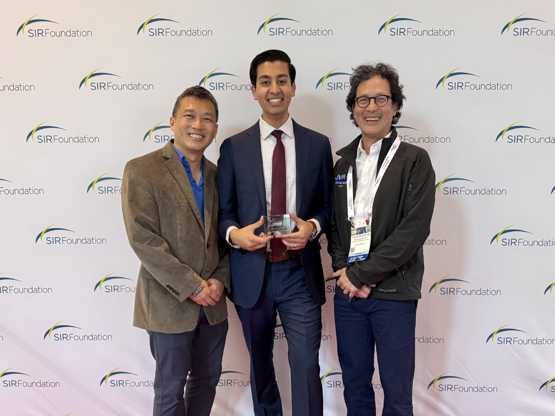
This article was informed by Dr. Short’s presentation at the SIR 2026 3rd Annual Quality and Performance Improvement (QPI) Division Forum, where he was awarded Best Presentation.
- Bukhari A, et al. Timeliness of Lung Cancer Diagnosis and Treatment. Fed Pract. 2017 Feb;34(Suppl 1):24S–29S)
- O'Hanlon C, et al. Comparing VA and Non-VA Quality of Care: A Systematic Review. J Gen Intern Med. 2017 Jan;32(1):105-121.
- Berger KI, Adam O, Dal Negro RW, Kaminsky DA, Shiner RJ, Burgos F, de Jongh FHC, Cohen I, Fredberg JJ. Validation of a Novel Compact System for the Measurement of Lung Volumes. Chest. 2021 Feb 2;159(6):2356-2365. doi: 10.1016/j.chest.2021.01.052
Related articles
-
IRQ Research
![]()
Inside Access: Iwan Paolucci, PhD
![]()
A new study investigating stereotactic thermal ablation of malignant liver tumors has found that the technology is likely to be safe, effective and reproducible in the U.S. healthcare system.
-
IRQ Research
![]()
Inside access: Masao Yamamoto-Ramos, MD, EBIR
![]()
Masao Yamamoto-Ramos, MD, EBIR, discusses how recognizing compensated venous phenotypes could support more personalized follow-up and treatment strategies in interventional radiology
Inside Access
Venous
-
IRQ Research
![]()
Inside access: Steve Landers MBBS
![]()
Inside the 6-month results of a prospective case series investigating transarterial embolization for plantar heel pain.
Inside Access
Pain Management
-
IRQ Research
![]()
How to get published in JVIR
![]()
Tips for standing out in a highly competitive submission pool
JVIR
IR research
-
IRQ Research
![]()
How to get started in research
![]()
A successful researcher doesn’t necessarily need a PhD—but they do need a varied support network, a strong plan and a commitment to persistence.
IR research
Research Funding: Awards
Research Funding: Grants
Women in IR
-
IRQ Research
Inside access: Aakash Gupta, MD
Dr. Aakash Gupta discusses TEPS as an effective last-resort option for portal decompression for specific patient populations.
JVIR
![]()
Related articles
![]()
-
IRQ Research
Inside Access: Iwan Paolucci, PhD
A new study investigating stereotactic thermal ablation of malignant liver tumors has found that the technology is likely to be safe, effective and reproducible in the U.S. healthcare system.
-
IRQ Research
Inside access: Masao Yamamoto-Ramos, MD, EBIR
Masao Yamamoto-Ramos, MD, EBIR, discusses how recognizing compensated venous phenotypes could support more personalized follow-up and treatment strategies in interventional radiology
Inside Access Venous -
IRQ Research
Inside access: Steve Landers MBBS
Inside the 6-month results of a prospective case series investigating transarterial embolization for plantar heel pain.
Inside Access Pain Management -
IRQ Research
How to get published in JVIR
Tips for standing out in a highly competitive submission pool
JVIR IR research -
IRQ Research
How to get started in research
A successful researcher doesn’t necessarily need a PhD—but they do need a varied support network, a strong plan and a commitment to persistence.
IR research Research Funding: Awards Research Funding: Grants Women in IR -
IRQ Research
Inside access: Aakash Gupta, MD
Dr. Aakash Gupta discusses TEPS as an effective last-resort option for portal decompression for specific patient populations.
JVIR
Related articles