
Inside Access
JVIR
Interventional Oncology
IR Quarterly
Inside access: Masao Yamamoto-Ramos, MD, EBIR
Understanding recurrence in pelvic venous disorders through the SVP classification
Yamamoto-Ramos M, et al. Understanding Recurrence in Pelvic Venous Disorders through the Symptoms-Varices-Pathophysiology Classification: A Multicenter Prospective Cohort Study. J Vasc Interv Radiol. 2026 Jul;37(7):108758.
Tell us about you, your team and your institution.
Masao Yamamoto-Ramos, MD, EBIR: I am an interventional radiologist and PhD candidate at the University of Zaragoza, Spain. This work was developed within the Minimally Invasive Techniques Research Group (GITMI), led by Professor Miguel Ángel de Gregorio and Professor José Andrés Guirola. Our group focuses on vascular and interventional radiology, image-guided therapies, and clinical outcomes research, with a particular interest in pelvic venous disorders.
This study forms part of my doctoral research program and represents one of the largest prospective multicenter cohorts evaluating recurrence after embolization for pelvic venous disorders, involving six centers across Spain operating under a unified diagnostic, treatment, and follow-up protocol.

Why did you pursue this research topic?
Dr. Yamamoto-Ramos: Clinical recurrence after embolization remains one of the most challenging and least understood aspects of pelvic venous disorders. Although technical success rates are generally high, a significant number of patients continue to experience recurrent symptoms.
We wanted to understand why this occurs and whether the SVP classification could help identify patients at higher risk. Our goal was to move beyond simply describing recurrence and instead better understand the hemodynamic mechanisms that drive it.
Professor Miguel Ángel de Gregorio: Pelvic venous disorders have been one of the main research interests of our Minimally Invasive Techniques Research Group (GITMI) at the University of Zaragoza for many years. For a long time, these patients remained underdiagnosed despite the availability of minimally invasive treatments capable of significantly improving their quality of life. Our group has contributed to the development and evaluation of systematic embolization of the four major pelvic venous axes. This study has now revealed a new challenge: understanding how post-embolization redistribution of venous flow through pre-existing escape pathways may explain part of the recurrence observed after technically successful procedures. A better understanding of these hemodynamic changes will be essential for developing increasingly personalized treatments and further improving patient outcomes.
What are the key takeaways from your research?
Dr. Yamamoto-Ramos: The most important finding was that V3b non-saphenous lower-extremity varices were the only independent predictor of recurrence after embolization. Patients with V3b pathways experienced recurrence earlier and more frequently than other patients.
These findings suggest that recurrence is strongly related to a compensated hemodynamic phenotype characterized by persistent extrapelvic decompression pathways rather than technical failure of the embolization itself.
More importantly, the study suggests that V3b varices are not simply an anatomic finding, but rather a marker of a compensated hemodynamic phenotype with distinct clinical behavior.
Figure 6. Evolution of a compensated pelvic venous disorder.
(A) Initial angiography shows a compensated pelvic venous disorder with multiple pelvic escape pathways.
(B) The four major pelvic venous axes were successfully embolized, while a small residual escape pathway remained untreated.
(C) During follow-up, venous flow redistributed through this pathway, leading to collateral enlargement and symptomatic recurrence despite technically successful embolization.
(D) Selective re-embolization of the residual escape pathway restored hemodynamic control, illustrating that recurrence may result from venous adaptation rather than treatment failure.


How might this research influence treatment, practice, or interventional radiology?
Dr. Yamamoto-Ramos: Our findings support a shift toward phenotype-driven management. The study suggests that patients with compensated phenotypes should not necessarily undergo more aggressive initial embolization but should receive closer surveillance and individualized follow-up.
Identifying V3b pathways before treatment may help physicians better counsel patients regarding recurrence risk and design more targeted follow-up strategies.
In practical terms, we believe V3b patients should be considered a higher-risk subgroup that may benefit from closer surveillance during the first months after embolization, when recurrence appears to be most common.
Did the authors identify whether recurrence in V3b patients was primarily driven by untreated escape pathways versus true recanalization after embolization?
Dr. Yamamoto-Ramos: Our findings suggest that recurrence was predominantly related to persistent or newly recruited escape pathways rather than recanalization of previously treated pelvic axes.
In most recurrent cases, the embolized ovarian and internal iliac venous trunks remained occluded, while symptoms were associated with hemodynamically active pelvic escape routes such as pudendal, obturator, gluteal or round ligament pathways.
This supports the concept that recurrence in V3b patients is mainly a consequence of a compensated venous phenotype with persistent redistribution of pelvic venous hypertension.
Should the presence of V3b varices change the initial embolization strategy or prompt more aggressive treatment of extrapelvic escape pathways upfront?
Dr. Yamamoto-Ramos: At present, we do not believe our data support routine escalation of the initial procedure.
Systematic embolization of the four major pelvic venous axes remains an effective first-line strategy. However, the presence of V3b varices should alert physicians that these patients represent a compensated phenotype with a higher risk of recurrence.
Rather than more aggressive upfront treatment, our results support closer follow-up and selective treatment of clinically relevant escape pathways when recurrence is demonstrated.
How reproducible was the SVP classification across centers, particularly for identifying hemodynamically significant V3b disease on duplex ultrasound and venography?
Dr. Yamamoto-Ramos: One of the strengths of our study is that all participating centers used a unified diagnostic, treatment, and follow-up protocol. Procedures were performed by experienced interventional radiologists using standardized imaging criteria and centralized data management.
This approach improved consistency in SVP classification and in the identification of V3b pathways. Nevertheless, further studies specifically evaluating interobserver agreement and reproducibility of the SVP classification would be valuable.
Any next steps or plans for follow-up research?
Dr. Yamamoto-Ramos: Absolutely. This study has opened several new research directions for our group.
One of the areas we are particularly excited about is the application of machine learning techniques to pelvic venous disorders. As multicenter datasets continue to grow, we believe these tools may help us better understand recurrence patterns and identify factors associated with different clinical outcomes.
More broadly, we remain interested in understanding how venous hemodynamics evolve after treatment and how different patient phenotypes behave over time. We hope that future research will allow us to move toward increasingly personalized approaches to follow-up and treatment planning in patients with pelvic venous disorders.
For us, this study is not the end of the story—it is really the beginning of a new way of thinking about recurrence in PeVD.
We hope this work encourages broader discussion about venous phenotyping and personalized treatment strategies in pelvic venous disorders.
Related articles
-
IRQ Research
![]()
Inside Access: Iwan Paolucci, PhD
![]()
A new study investigating stereotactic thermal ablation of malignant liver tumors has found that the technology is likely to be safe, effective and reproducible in the U.S. healthcare system.
-
IRQ Research
![]()
Inside access: Masao Yamamoto-Ramos, MD, EBIR
![]()
Masao Yamamoto-Ramos, MD, EBIR, discusses how recognizing compensated venous phenotypes could support more personalized follow-up and treatment strategies in interventional radiology
Inside Access
Venous
-
IRQ Research
![]()
Inside access: Steve Landers MBBS
![]()
Inside the 6-month results of a prospective case series investigating transarterial embolization for plantar heel pain.
Inside Access
Pain Management
-
IRQ Research
![]()
How to get published in JVIR
![]()
Tips for standing out in a highly competitive submission pool
JVIR
IR research
-
IRQ Research
![]()
How to get started in research
![]()
A successful researcher doesn’t necessarily need a PhD—but they do need a varied support network, a strong plan and a commitment to persistence.
IR research
Research Funding: Awards
Research Funding: Grants
Women in IR
-
IRQ Research
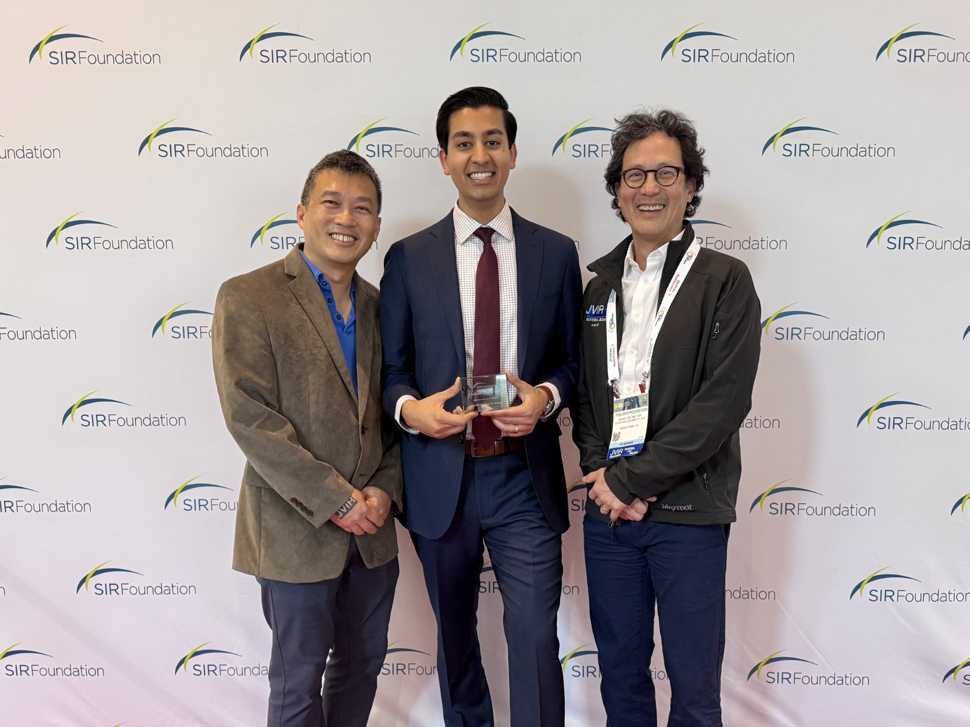
Inside access: Aakash Gupta, MD
Dr. Aakash Gupta discusses TEPS as an effective last-resort option for portal decompression for specific patient populations.
JVIR
![]()
Related articles
![]()
-
IRQ Research
Inside Access: Iwan Paolucci, PhD
A new study investigating stereotactic thermal ablation of malignant liver tumors has found that the technology is likely to be safe, effective and reproducible in the U.S. healthcare system.
-
IRQ Research
Inside access: Masao Yamamoto-Ramos, MD, EBIR
Masao Yamamoto-Ramos, MD, EBIR, discusses how recognizing compensated venous phenotypes could support more personalized follow-up and treatment strategies in interventional radiology
Inside Access Venous -
IRQ Research
Inside access: Steve Landers MBBS
Inside the 6-month results of a prospective case series investigating transarterial embolization for plantar heel pain.
Inside Access Pain Management -
IRQ Research
How to get published in JVIR
Tips for standing out in a highly competitive submission pool
JVIR IR research -
IRQ Research
How to get started in research
A successful researcher doesn’t necessarily need a PhD—but they do need a varied support network, a strong plan and a commitment to persistence.
IR research Research Funding: Awards Research Funding: Grants Women in IR -
IRQ Research
Inside access: Aakash Gupta, MD
Dr. Aakash Gupta discusses TEPS as an effective last-resort option for portal decompression for specific patient populations.
JVIR
Related articles