
Inside Access
JVIR
Interventional Oncology
IR Quarterly
Inside Access: Iwan Paolucci, PhD
Stereotactic thermal ablation of malignant liver tumors
Paolucci I, et al. Stereotactic Percutaneous Thermal Ablation of Primary and Metastatic Liver Tumors: A Prospective Registry-Based Study. JVIR, 2026;37.
Tell us about you, your team and your institution.
Iwan Paolucci, PhD: The team for this study is very interdisciplinary, with investigators across the departments of interventional radiology, anesthesia and perioperative medicine, and imaging physics here at UT MD Anderson. In addition, we had Professor Reto Bale from the University of Innsbruck, Austria, who has performed stereotactic liver ablation for over two decades, as a mentor throughout the process. Our department performs over 300 liver ablation procedures per year with a steady year-over-year growth of almost 20% over the past decade. My group, along with fellow author Bruno C. Odisio, MD, FSIR, heavily focuses on research around thermal ablation of liver tumors.
Why did you pursue this topic?
Dr. Paolucci: I did my MSc and PhD on stereotactic guidance for ablation and resection of liver tumors. I worked on such projects for over 5 years at the University of Bern, Switzerland, before joining UT MD Anderson. It remains a topic I am very passionate about and teach to faculty, residents and students. Dr. Odisio, who was my postdoctoral supervisor, became interested in this technology after he met Prof. Bale and learned about his technique and excellent outcomes.
Naturally, we decided to start this program at UT MD Anderson. Since it was the first large-scale application in the U.S., we chose to pilot the program under a study and prospectively collect accuracy data, timing and user feedback surveys. Timing and accuracy were important metrics that we monitor continuously to make sure the technology meets our expectations. It allowed us to rely on data rather than subjective feedback—especially with so many IR faculty involved. Long story short, we chose to use a scientific approach in a product trial which eventually benefited both the product evaluation and our research endeavors.
What are the key takeaways from your research?
Dr. Paolucci: The main takeaway is that the results of this technology are reproducible in the U.S. healthcare system. Safety and accuracy are comparable to many European studies and short-term oncologic outcomes are better than many reports on conventional procedures. Since it was a histology agnostic study, this must be interpreted accordingly, and local tumor progression rates should be interpreted cautiously. The study also shows that the introduction of this technology is a collaborative effort and requires good communication with the anesthesia team and imaging physicists. In addition, it provides data that favors high frequency jet ventilation (HFJV) for respiratory motion control.
For someone unfamiliar with stereotactic thermal ablation, can you briefly explain how this technique works and what makes it different from traditional liver tumor ablation methods?
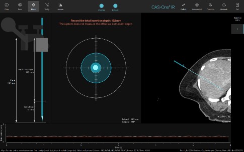
Dr. Paolucci: A stereotactic system continuously measures the location and orientation of your instruments (in this case a mechanical arm) and the patient—typically using a stereo infrared camera. It also uses patient-to-image registration to establish the spatial relationship between the CT image and the patient. As such it can compute the location of the physical instruments on the image at any time. We then plan the ablation applicator trajectory on the CT image, and the system helps the IR align the aiming device using a crosshair viewer or other visualization techniques. The same applies to robotic guidance—with the only difference that the alignment is done by the robot using the actuators in the arm. Once aligned, the IR can then insert the ablation applicator through the guidance channel on the aiming device which now points exactly into the direction that was planned on the CT. The applicator can be inserted directly into the target or a bit shorter in case of critical structures without intermittent imaging. After the placement, another CT scan is acquired and fused on the system to confirm that the applicator is placed as planned and manual corrections are made as necessary based on the system output. Newer versions also have ablation confirmation available to assess the margins after the ablation.
Compared to traditional liver ablation, this technique provides a high level of standardization and reproducibility through detailed planning, guidance and verification. A more junior physician can achieve similar results to a senior physician faster since it removes some of the user dependency—especially in complex cases.
Your study compares breath-hold techniques with HFJV. What did you find regarding targeting accuracy, and why do you think HFJV performed better?
Dr. Paolucci: We found that using jet ventilation resulted in better targeting accuracy compared to breath holds. It is most likely due to better reproducibility. Using breath holds, there is more variability in the final lung volumes at the end of breath holds. This occurs if the team does not wait long enough for full expiration or if ventilator settings are not set properly. With communication this can usually be optimized, however these inconsistencies can be completely avoided by nonstop jet ventilation.
Based on your results, how safe and effective do you think this approach is for routine use in U.S. clinical practice, and are there any patients who might benefit most from it?
Dr. Paolucci: Overall, our data shows that the technique is safe and effective. Major adverse events are not increased compared to conventional ablations and short-term local outcomes are better than what has been reported historically. These are encouraging results given that the learning curve of this methodology during its initial implementation does not seem to affect safety, while keeping outcomes within the range reported by our group previously.
How might this research influence treatment, practice or clinical processes in interventional radiology?
Dr. Paolucci: Interventional radiology has become an integral component of multidisciplinary cancer care, offering therapies that can meaningfully improve patient outcomes and quality of life. Despite these advances, our specialty continues to face challenges related to the standardization of patient selection, procedural techniques, and outcome assessment, all of which are necessary to enable broader adoption and scalability across institutions and practice settings.
This research speaks to two important opportunities for the field. First, it reinforces the importance of evaluating innovations within the framework of well-designed clinical investigations. By doing so, we can rigorously assess efficacy and safety and generate much-needed evidence to establish best practices that demonstrate the value of interventional oncology therapies. Second, the study addresses a critical barrier to the broader adoption of liver ablation: the ability to standardize and disseminate high-quality procedural techniques. Establishing reproducible methodologies, objective quality metrics and scalable training models is essential to ensure that patients, regardless of geographic location, can benefit from consistently safe and effective treatments.
More broadly, this work reflects the ongoing evolution of interventional radiology toward a more evidence-based, data-driven and outcomes-focused specialty. By combining innovation with rigorous clinical evaluation and reproducible procedural standards, we can accelerate the acceptance and adoption of our therapies, strengthen our role within multidisciplinary oncology teams and expand access to high-quality cancer care for patients worldwide.
Any next steps or plans for follow‑up research?
Dr. Paolucci: We finished patient accrual (N=50) on the STEREOLAB study (NCT05361551) in October 2025 and are now awaiting 2-years of follow-up. This study combined stereotactic guidance, CT during hepatic arteriography and ablation confirmation using biomechanical deformable image registration and AI-based segmentation.
Right now, this system is available in the department and IRs are using it regularly. Once we reach a larger number of patients with at least 2 years of follow-up, we will revisit the data and look at longer term oncologic outcomes, especially the newer versions with improved AI-based ablation confirmation. We are also exploring other applications than liver ablation.
From the anesthesia side, we have additional research projects investigating the use of HFJV during these procedures irrespective of using stereotactic guidance. This technique is still rather new for us and there is a lot to learn and optimize. Initially, we used HFJV only for stereotactic liver ablations, but we increasingly use it for conventional procedures as well.
Related articles
-
IRQ Research
![]()
Inside Access: Iwan Paolucci, PhD
![]()
A new study investigating stereotactic thermal ablation of malignant liver tumors has found that the technology is likely to be safe, effective and reproducible in the U.S. healthcare system.
-
IRQ Research
![]()
Inside access: Masao Yamamoto-Ramos, MD, EBIR
![]()
Masao Yamamoto-Ramos, MD, EBIR, discusses how recognizing compensated venous phenotypes could support more personalized follow-up and treatment strategies in interventional radiology
Inside Access
Venous
-
IRQ Research
![]()
Inside access: Steve Landers MBBS
![]()
Inside the 6-month results of a prospective case series investigating transarterial embolization for plantar heel pain.
Inside Access
Pain Management
-
IRQ Research
![]()
How to get published in JVIR
![]()
Tips for standing out in a highly competitive submission pool
JVIR
IR research
-
IRQ Research
![]()
How to get started in research
![]()
A successful researcher doesn’t necessarily need a PhD—but they do need a varied support network, a strong plan and a commitment to persistence.
IR research
Research Funding: Awards
Research Funding: Grants
Women in IR
-
IRQ Research
Inside access: Aakash Gupta, MD
Dr. Aakash Gupta discusses TEPS as an effective last-resort option for portal decompression for specific patient populations.
JVIR
![]()
Related articles
![]()
-
IRQ Research
Inside Access: Iwan Paolucci, PhD
A new study investigating stereotactic thermal ablation of malignant liver tumors has found that the technology is likely to be safe, effective and reproducible in the U.S. healthcare system.
-
IRQ Research
Inside access: Masao Yamamoto-Ramos, MD, EBIR
Masao Yamamoto-Ramos, MD, EBIR, discusses how recognizing compensated venous phenotypes could support more personalized follow-up and treatment strategies in interventional radiology
Inside Access Venous -
IRQ Research
Inside access: Steve Landers MBBS
Inside the 6-month results of a prospective case series investigating transarterial embolization for plantar heel pain.
Inside Access Pain Management -
IRQ Research
How to get published in JVIR
Tips for standing out in a highly competitive submission pool
JVIR IR research -
IRQ Research
How to get started in research
A successful researcher doesn’t necessarily need a PhD—but they do need a varied support network, a strong plan and a commitment to persistence.
IR research Research Funding: Awards Research Funding: Grants Women in IR -
IRQ Research
Inside access: Aakash Gupta, MD
Dr. Aakash Gupta discusses TEPS as an effective last-resort option for portal decompression for specific patient populations.
JVIR
Related articles